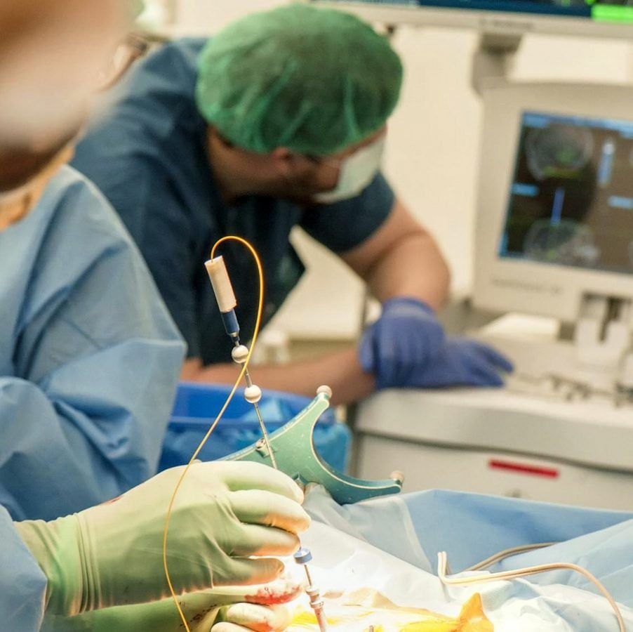
A ‘smart needle’ with a camera that makes brain surgery safe has been used on patients for the first time.
The device – as thin as a human hair – enables surgeons to avoid piercing vessels that could cause a life-threatening bleed.
It detects them with up to 98 percent accuracy by lighting up risky areas with infra-red light.
Professor Robert McLaughlin, chair of biophotonics at Adelaide University’s Medical School, said: “Brain biopsies are a common procedure carried out to diagnose brain tumour and other diseases.
“It’s a minimally invasive operation – but still carries the risk of damage to blood vessels that is potentially fatal.
“The imaging needle lets surgeons ‘see’ at-risk blood vessels as they insert the needle – allowing them to avoid causing bleeds.
“The fibre-optic camera – the size of a human hair – shines infrared light onto the brain tissue.
“And the computer system behind the needle identifies the blood vessel and alerts the surgeon.”
Currently, about one-in-a-hundred patients will die following the procedure – with up to three times as many left disabled.
The imaging needle has been successfully used in initial trials involving 11 patients at Sir Charles Gairdner Hospital in Perth, Western Australia.
Prof McLaughlin said: “These patients were undergoing other types of neurosurgery and consented to allow us to safely test how well the imaging needle was able to detect blood vessels during surgery.
“This is the first reported use of such a probe in the human brain during live surgery – and is the first step in the long process required to bring new tools like this into clinical practice.”
He is hoping it could be in widespread clinical use within the next five years.
The study published in Science Advances says the tiny needle has a fibre-optic camera encased inside.
Trial leader Prof Christopher Lind, consultant neurosurgeon at Sir Charles Gairdner Hospital and the University of Western Australia, said: “Bleeds are a risk in many types of neurosurgery.
“There is a great opportunity for new technologies like this to help us reduce those risks.
“To have a tool that can see blood vessels as we proceed through the brain would revolutionise neurosurgery.
“It will open the way for safer surgery – allowing us to do things we’ve not been able to do before.”
A brain biopsy is a procedure to remove a sample of abnormal tissue for examination under a microscope. The cells taken can show if it is benign or cancerous.
Needles are used to access tumours or lesions that are deeper in the brain. A neurosurgeon performs the biopsy surgery to obtain brain tissue samples.
A physician called a neuropathologist examines the sample cells and performs a variety of tests to make a diagnosis of the tumor type and grade. This process takes 5 to 7 days before final results are known.

